An overview of recent realisations
Epilog is a spin-off of HyCT’s MEDISIP group founded in 2016, focused around the idea that by using advanced signal analysis techniques, EEG can be exploited to its fullest potential and become a window into the brain. Enabling advanced EEG processing and making it accessible to clinical practice is Epilog’s driving force.
Read moreMOLECUBES is a spin-off from HyCT’s MEDISIP group, focusing on preclinical benchtop scanners. Imaging techniques such as computed tomography (CT), positron emission tomography (PET) and single photon emission computed technology (SPECT) are widely used to investigate drug development, as they provide anatomical and functional 3D information about in vivo biological processes (i.e. in living plants or small animals).
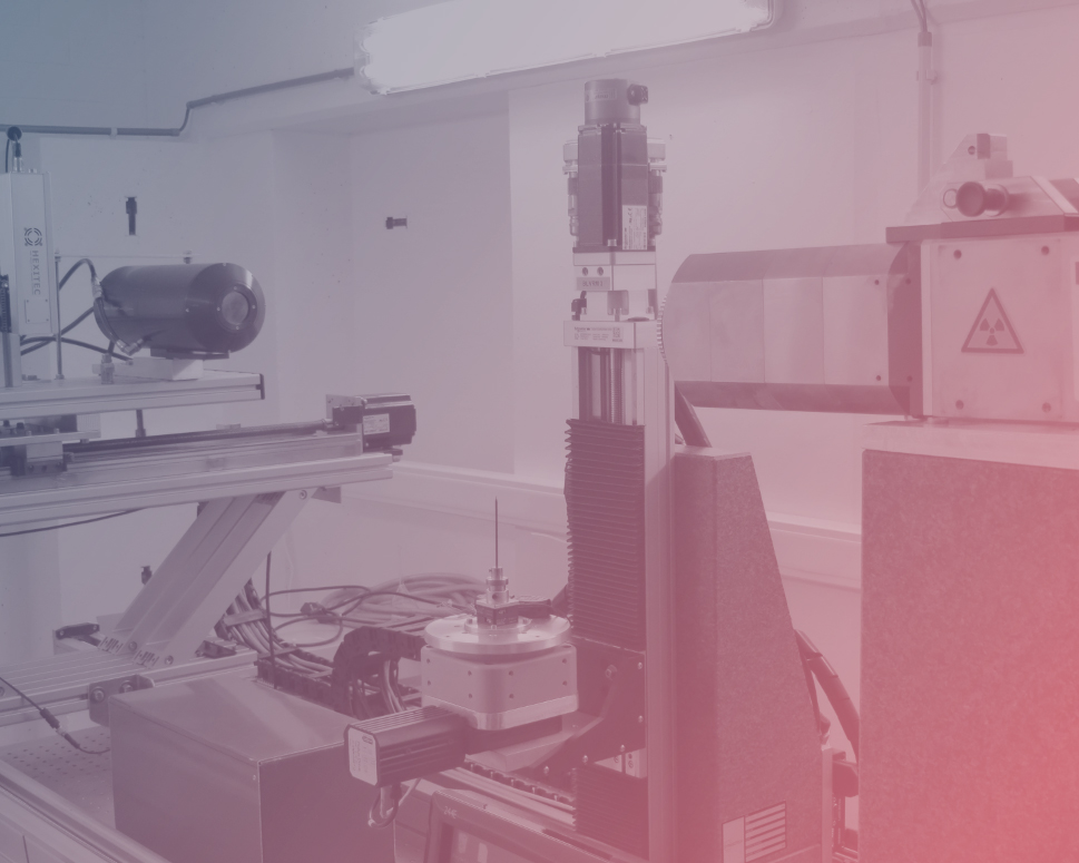
READ moreThe Centre for X-ray Tomography (UGCT) is the UGent Expertise Centre and core facility performing research on and with the X-ray micro-CT technique, and founding father of the HyCT consortium. UGCT also acts as a user facility that offers the use of X-ray CT to researchers of many scientific domains in their research. It was founded in 2006 and is one of the few CT centers in the world that cover the complete CT workflow: physics and instrumentation, data reconstruction and data analysis.
READ moreHaving co-designed all dedicated scanners in the UGCT lab, a group of researchers of the UGCT core saw an opportunity to launch their own service (Inside Matters) and hardware (XRE) spin-offs in 2007 and 2011 respectively. Since then they have been continuously pushing the boundaries of 3D and 4D CT imaging from a detector side as well as on reconstruction and post-processing.
READ MOREFEops, a spin-off of HyCT’s IBITECH research group, offers the first and only patient-specific simulation technology for structural heart interventions, surpassing basic anatomical measurement by accurately predicting how devices will interact with each unique patient. Their end-to-end solution adds value at every step in the structural heart product life cycle, helping manufacturers accelerate time to market and offering insights that help physicians achieve better patient outcomes.
READ MOREThe Sphynx project, run by HyCT’s MEDISIP group, will build the first Total body time-of-flight PET (TB-TOF-PET) scanner, with significantly improved technical capabilities, to be deployed as a research tool to study physiology in plants, large animals and humans.
READ MOREEpilog is a spin-off of HyCT’s MEDISIP group founded in 2016, focused around the idea that by using advanced signal analysis techniques, EEG can be exploited to its fullest potential and become a window into the brain. Enabling advanced EEG processing and making it accessible to clinical practice is Epilog’s driving force.
Bringing advanced EEG technology into clinical practice is a complex challenge. Several advanced signal and image processing techniques have been studied and validated in epilepsy. Unfortunately, despite very promising results of validation studies, they have not been widely disseminated in clinical practice. Epilog believes this is due to the technological barriers that exist for clinical centers who do not have resources that can be dedicated to this task. The core philosophy of Epilog is to bring advanced EEG diagnostic technology to clinical practice as a service. A service that allows EEG readers to focus on what really matters: the interpretation of the data to the benefit of the patient or the study. Epilog offers interpretation guidelines, training and symposia to continuously involve and train our collaborating centers, clinicians and industry. Their current services are focused on epilepsy. They offer 3 different solutions.
Epilog PreOp provides an efficient analysis of long-term EEG recordings and HD-EEG recordings in the preoperative work-up, including electrical source imaging for the localization of the irritative zone. Epilog DX is focused on making the analysis of 24 hours or overnight EEG recordings more approachable and less time consuming. Our services for industry are dedicated to clinical trials, offering our biomarkers to de-risk them.
The basis of all these technologies, as well as patented IP behind this, was developed over a 10 year span within the HyCT’s MEDISIP core group in close relationship to Ghent University Hospital.
MOLECUBES is a spin-off from HyCT’s MEDISIP group, focusing on preclinical benchtop scanners. Imaging techniques such as computed tomography (CT), positron emission tomography (PET) and single photon emission computed technology (SPECT) are widely used to investigate drug development, as they provide anatomical and functional 3D information about in vivo biological processes (i.e. in living plants or small animals).
While the effectiveness of the current generation of scanners to monitor the effect of new medications remains undisputed, they are bulky, very costly to deploy, maintain and operate – and thus only available to a limited amount of researchers.
That is why MOLECUBES, after about 5 years of HyCT system development resulting in 3 patents and numerous software routines, end of 2015 launched a more easy and flexible approach to in vivo scanning of lab animals in the life science research industry. Bringing to market the world’s smallest desktop PET, SPECT and CT scanner family for the 3D imaging of small animals.
In SPECT, patented lofthole technology and laser sintered collimators combined with high-resolution detectors result in a high-end true benchtop imager. In-house developed image reconstruction software guarantees fast imaging and excellent image quality. All common SPECT-labeled therapeutic and diagnostic imaging tracers can be imaged.
In PET, sub-millimeter image resolution is achieved through the combination of monolithic scintillators, the latest photon counting technology and GPU-based event positioning and iterative image reconstruction. The 5-ring configuration ensures best-in-class sensitivity over a field-of-view adequate for whole-body mouse and rat imaging at high count rate. In-house hardware allows for dynamic and gated studies.Developing and selling its desktop imaging systems for over 5 years now, MOLECUBES is becoming a worldwide player in the preclinical imaging business by selling its products to academic, government and private biomedical research laboratories, pharmaceutical and biotechnology companies and contract research organizations (CRO).
The Centre for X-ray Tomography (UGCT) is the UGent Expertise Centre and core facility performing research on and with the X-ray micro-CT technique, and founding father of the HyCT consortium. UGCT also acts as a user facility that offers the use of X-ray CT to researchers of many scientific domains in their research. It was founded in 2006 and is one of the few CT centers in the world that cover the complete CT workflow: physics and instrumentation, data reconstruction and data analysis.
The UGCT is operated by a multi-disciplinary team and is currently an interfactultary collaboration between 3 research groups: the Radiation Physics group, the Pore-Scale Processes in Geomaterials Research group and the Laboratory for Wood technology.
UGCT performs research on laboratory based very high-resolution X-ray tomography and its applications, covering the complete imaging chain. This includes CT scanner development, simulation of the imaging chain, the study of novel imaging techniques, developing tomographic reconstruction methods (including iterative reconstruction techniques and GPU-based code) and 3D image analysis methods. The developed hardware and methodology are being used within UGCT for geological and wood technology research.
As a user facility, UGCT offers external research groups and companies access to its unique combination of state-of-the-art in-house developed high-resolution X-ray CT systems and its CT expertise. The flexibility of the systems and the knowledge of the researchers involved allows to perform challenging experiments in various research fields, continuously pushing the limits of what is currently possible with CT.
Curious about what they can do? Visist their website and have a look at some examples of CT-scans.
Having co-designed all dedicated scanners in the UGCT lab, a group of researchers of the UGCT core saw an opportunity to launch their own service (Inside Matters) and hardware (XRE) spin-offs in 2007 and 2011 respectively. Since then they have been continuously pushing the boundaries of 3D and 4D CT imaging from a detector side as well as on reconstruction and post-processing.
Having co-designed all dedicated scanners in the UGCT lab, a group of researchers of the UGCT core saw an opportunity to launch their own service (Inside Matters) and hardware (XRE) spin-offs in 2007 and 2011 respectively. Since then they have been continuously pushing the boundaries of 3D and 4D CT imaging from a detector side as well as on reconstruction and post-processing. Software packages like OCTOPUS and Morpho+, initially designed for academic use only, were slowly but surely commercialized until their own “TOM” scanline is now emerging.
Inside Matters and XRE engineering, today merged and acquired by TESCAN, are 2 HyCT spin-offs from the UGCT core group. With expertise built around customized high resolution CT service provision as well as X-ray and CT-system design both spin-offs make their name in the field of ex-vivo X-ray imaning. Combining forces with TESCAN, there are now pioneering the emerging field of dynamic CT in the laboratory and offers innovative micro-CT systems that facilitate a diverse range of applications. Their micro-CT systems – CoreTOM, DynaTOM, and UniTOM XL – are designed for high throughput and flexibility, offering novel dynamic CT workflows that enable a host of in situ and 4D applications, from Earth Science to Materials Science and beyond.
FEops’ simulation technology uses advanced computational modeling and simulation, initiating from HyCT’s IBITECH lab, to provide manufacturers and physicians with pre-operative insights into the interaction between patient anatomy and heart implants. One example is FEops HEARTguide™. It is available on the European and Canadian market with indication for use in TAVI and LAAo. Outside Europe and Canada, FEops HEARTguide™ is only available for non-clinical use.
By combining routine preoperative CT images with patient-specific CM&S, FEops HEARTguide™ predicts the interaction between the TAVI device and the patient’s unique anatomy, including post-implantation deformation, allowing physicians to assess the risk for aortic regurgitation and conduction abnormalities. Considering the extension of TAVI indications to younger, lower-risk patients (of which many have bicuspid aortic valves), these insights are key. Among these patients, the chance of complications such as paravalvular regurgitation and new permanent pacemaker remains high. This illustrates the need to more effectively risk-stratify bicuspid and lower risk aortic valve patients.
Using FEops HEARTguide™ in bicuspid aortic valve cases gives physicians detailed preoperative information about the interaction between the replacement valve and the patient’s anatomy – helping them improve outcomes and minimize risk.
The Sphynx project, run by HyCT’s MEDISIP group, will build the first Total body time-of-flight PET (TB-TOF-PET) scanner, with significantly improved technical capabilities, to be deployed as a research tool to study physiology in plants, large animals and humans.
Where current clinical PET devices have 3 to 5 rings of detectors (plus a moveable bed) the Sphynx concept is to concatenate and integrate 20 or more detector rings, to obtain a long detector cylinder. The device will be large enough to house large subjects for single-shot imaging, will have a 20-fold improved sensitivity, fourfold longer axial field of view, and twofold better spatial resolution (compared to state of the art clinical PET).
It is supported by a large scientific Flemish community of engineers, pharmacists, bioengineers, clinical and veterinary specialists The specific domains studied within human, animal, and plant physiology are neurology, oncology, endocrinology, gastroenterology, paediatrics, and plant ecophysiology. Results from Sphynx may lead to new clinical therapies (drugs, cellular therapy, radiotherapy, surgical procedures, transplantation strategies, …)and solutions for agriculture, forestry and ecosystem management.
Sphynx has kicked off in 2020 with both local, governemtal and European support and aims at having a first TOF one-ring demonstrator ready by the end of 2021. All groups related to TB TOF are invited to contact us to see where and how collaborations is possible.